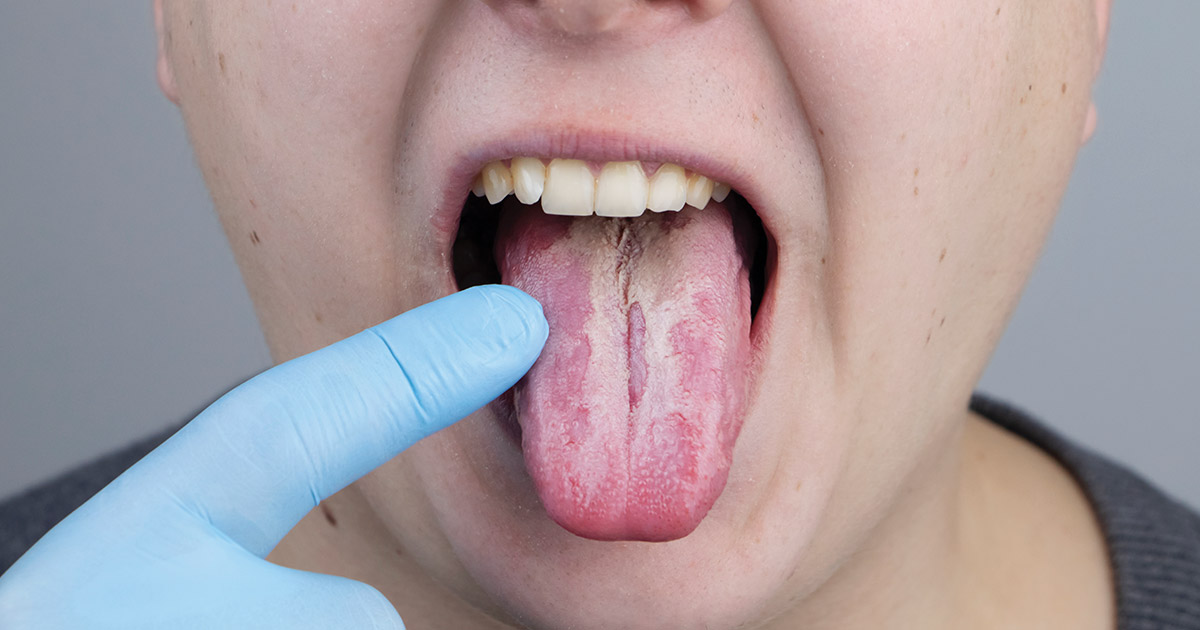
Red and white lesions in the oral cavity from a clinical perspective
The oral mucosa has many white lesions that are benign and do not require treatment
Post by DR. GOUNSIA AMIN on Saturday, December 23, 2023

DIAGNOSIS AND TREATMENT
Introduction
There are four groups of oral lesions that can be classified as ulcerations, pigmentations, exophytic lesions, and red – white lesions. White and red patches of the oral mucosa comprise of a significant group of disorders that arise from a diverse spectrum that ranges from traumatic lesions, infectious diseases, systemic and local immune mediated lesions, to potentially malignant disorders or a neoplasm. Oral cavity is most affected by the red and white lesions. To differentiate the various colours of the lesions in the oral cavity, one must know, the colour of the oral mucosa depends on the degree of keratinization, dilation, and concentration of blood vessels, the amount of melanin pigment, and the thickness of the epithelium.
The whiteness of the oral mucosa can be due to acanthosis, hyperkeratosis, and necrosis in the oral epithelium, vascularity reducing in the underlying lamina propria, and accumulation of fluid intracellular and extracellular in the epithelium. The red is caused by the enlarged blood vessels, atrophic epithelium and an increase in vascularisation and decrease in the number of epithelial cells. The combination of red and white lesions suggests an irregular epithelial surface that may be caused by a variety of processes, including chronic trauma, inflammation, and neoplasia.
Causes
The whiteness of oral mucosa is caused by:
- Keratinization of normally non-keratinized mucosa is one of the changes that occur in the epithelium.
- The normal keratinization of the mucosa has been increased.
- The epithelium has abnormal keratinization.
- Thickening of the epithelium, and epithelial edema.
- Fibrosis and decrease in the vascularity of the underlying mucosa, may also lead to the whiteness. It tends to appear deep without evident surface alterations.
The redness of oral mucosa is caused by:
- Vasodilation: The diameter of the blood vessels gets increased due to inflammation, but also possible with neoplasia.
- Vascular proliferation: The increase in the number of blood vessel occur due to the release of growth factor such as, vascular endothelial growth factor (VEGF), during inflammation and as a part of neoplasia.
- Leaky blood vessels (bleeding into tissues): It occurs due to trauma, occasionally due to an immune response.
- Epithelial thinning (atrophy and or reduced epithelial keratinization): It is due to abnormal cell turnover during normal healing, in response to trauma, or as a part of dysplasia.
Signs
- Colour: homogeneous red (erythroplakia) or mixed red and white (erythroleukoplakia); may appear lacy.
- Surface anatomy: flat, elevated, mass, ulceration.
- Surface texture: smooth, velvety, verrucous, or ulcerated.
- Consistency: Indurated or non-indurated.
- Tissue fragility.
- Paresthesia, numbness, discomfort, sensitivity to spicy/acidic foods.
- Nonhealing ulcer present for more than 2 weeks.
- Highest intraoral risk sites for neoplasia are lateral and ventral surfaces of the tongue, floor of mouth and retromolar area. Waldeyer tonsillar ring (tonsils, base of tongue) at increased risk.
Location
Single site, multiple sites, bilateral or unilateral presentation. Malignancies are less commonly bilateral.
Symptoms
Pain severity is variable in potential precancerous or malignant lesions: there may be no pain, some discomfort, aching or spontaneous stabbing pain. Throbbing pain or sensitivity to touch suggests possible underlying inflammation. Burning, stabbing sensation: suggests possible neural involvement and in combination with red or red-white lesions, malignancy must be considered.
Management
Despite a thorough clinical evaluation, some white lesions remain clinical problems despite being diagnosed. If the initial evaluation indicates that the white lesion is caused by trauma, it is essential to eliminate any possible irritants and re-examine the patient within ten days. If there is no significant clinical improvement, it is important to have an immediate biopsy to rule out a potential neoplastic change.
The oral mucosa has many white lesions that are benign and do not require treatment. Conditions that are inherited or developed, such as leukoedema, white sponge nevus, keratosis follicularis, hereditary benign intraepithelial dyskeratosis, and Fordyce granules, are included. Skin and mucosal grafts, Materia Alba associated with the gingiva or tongue, and keratotic lesions like hairy tongue are other benign conditions that don't require intervention.
Hypersensitivity to drugs, foods, or dental materials (such as denture adhesives, toothpastes, and mouth rinses) can result in red lesions that can appear anywhere in the oral cavity. Discontinuation of the offending substance is the main part of treatment and up to 40 mg/d of prednisone can promote healing. Traumatized vascular lesions may necessitate surgery or embolization to control bleeding. The diagnosis of pyogenic Granuloma and peripheral giant cell granuloma necessitates biopsy due to their resemblance to amelanotic melanoma.
Advice
If lesions are suspicious for malignancy: refer to a specialist with experience in mucosal disease and oncology, such as specialists in oral medicine, pathology, surgery, otorhinolaryngology, or oncology. May require biopsy with appropriate biopsy site selection, careful technique, special tissue stains and submission to pathologist experienced in oral mucosal disease. If the pathology report is not consistent with history and clinical presentation of the lesion, consider repeating pathologic review, repeat biopsy or refer. Preneoplastic lesions may evolve over time, so repeated biopsies may be needed during follow-up. Treatment is variable and largely dependent on diagnosis.
(Dr Gounsia Amin, Oral Medicine and Radiology. Email id: gousiaamin786@gmail.com)